25th UK Conference on
Medical Image Understanding and Analysis
COVID-19 Update
Due to future uncertainty caused by the COVID-19 pandemic, we have regretfully decided to cancel the physical meeting this year and move MIUA 2021 to a virtual context. The safety and well-being of MIUA participants is our top priority, therefore we believe that fully virtual meeting is the only viable option to run this conference smoothly.
As MIUA 2021 goes virtual this year, we will be using a virtual events platform across all three days, which will enable all of us to meet virtually, present the accepted papers and moderate live question and answer sessions, and deliver associated seminars and tutorials. The detailed schedule and guidelines about presentation will be released in due course. The conference registration fee will be reduced to reflect this change.
We appreciate the work that has gone into preparing contributions, hence we are still welcoming paper submissions until 28th March 2021 using our online submission system. Please note that this deadline is final and no further extensions can be granted at this point. All submissions will be peer-reviewed and accepted articles will be published as MIUA Proceedings by the Springer Publishing Group.
Thank you for your understanding, and we look forward to delivering MIUA 2021 in a way that is rewarding for our authors and interesting for the research community as a whole.
Scope
MIUA is a UK-based international conference for the communication of image processing and analysis research and its application to medical imaging and biomedicine. This is a rapidly growing subject with ever increasing real-world applicability.
MIUA welcomes all researchers in medical imaging including mathematicians, computer scientists, bioinformaticians, clinicians, engineers and bioscientists.
MIUA is the principal UK forum for communicating research progress within the community interested in image analysis applied to medicine and related biological science. The meeting is designed for the dissemination and discussion of research in medical image understanding and analysis, and aims to encourage the growth and raise the profile of this multi-disciplinary field by bringing together the various communities including among others:
| Brain imaging | Cancer | Cardiac Imaging | Circulation and Microcirculation |
| Computational anatomy and physiology | Computed Tomography | Dermatology | Imaging Physics |
| In-vivo intravital imaging | Inflammation | Magnetic Resonance Imaging | Microscopy | Neurology | Novel Imaging Methods | Ophthalmology | Optical Imaging |
| Positron Emission Imaging | Radiology | Tissue Perfusion | Ultrasound |
Keynote Speakers
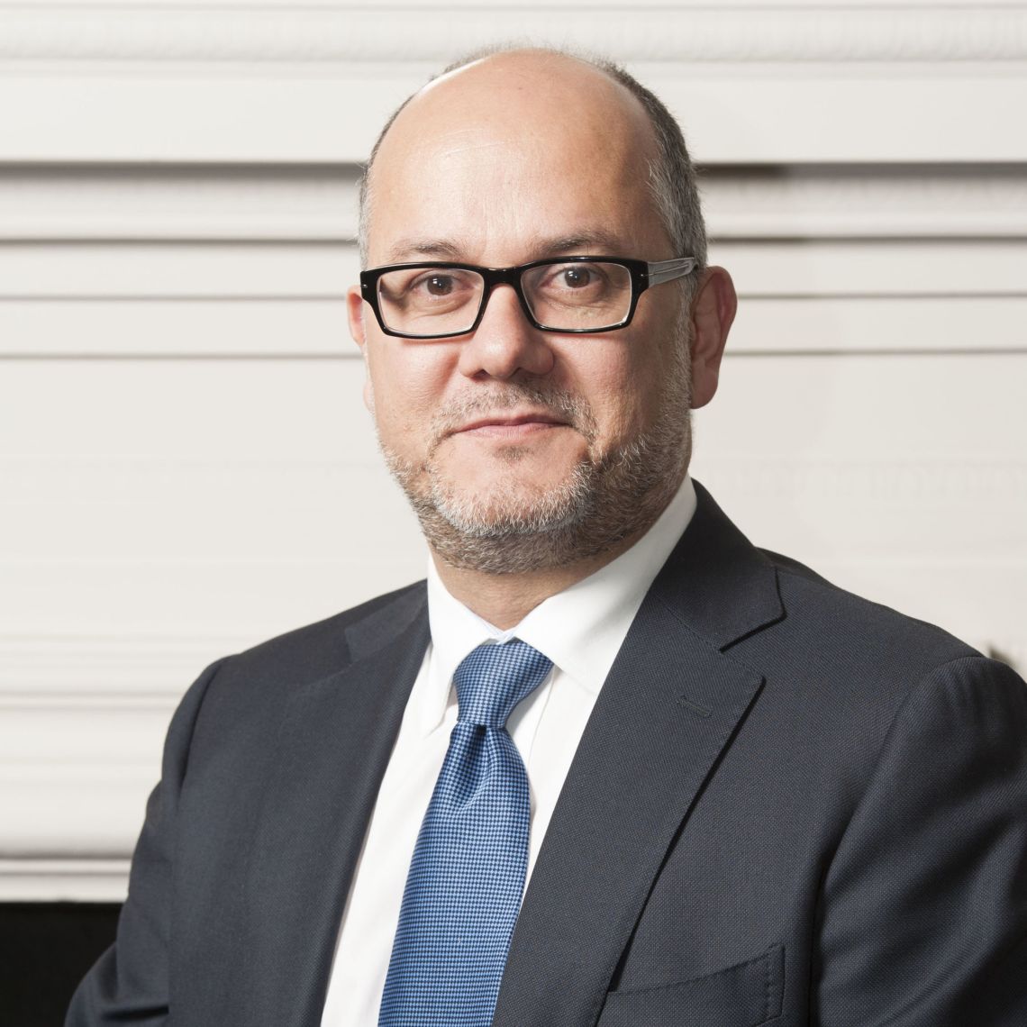
Prof. Mihaela van der Schaar
The John Humphrey Plummer Professor of Machine Learning, Artificial Intelligence and Medicine at the University of Cambridge
Prof. Mihaela van der Schaar
The John Humphrey Plummer Professor of Machine Learning, Artificial Intelligence and Medicine at the University of Cambridge
Biography: Prof. Mihaela van der Schaar is the John Humphrey Plummer Professor of Machine Learning, Artificial Intelligence and Medicine at the University of Cambridge, a Fellow at The Alan Turing Institute in London, and a Chancellor’s Professor at UCLA.
Mihaela was elected IEEE Fellow in 2009. She has received numerous awards, including the Oon Prize on Preventative Medicine from the University of Cambridge (2018), a National Science Foundation CAREER Award (2004), 3 IBM Faculty Awards, the IBM Exploratory Stream Analytics Innovation Award, the Philips Make a Difference Award and several best paper awards, including the IEEE Darlington Award.
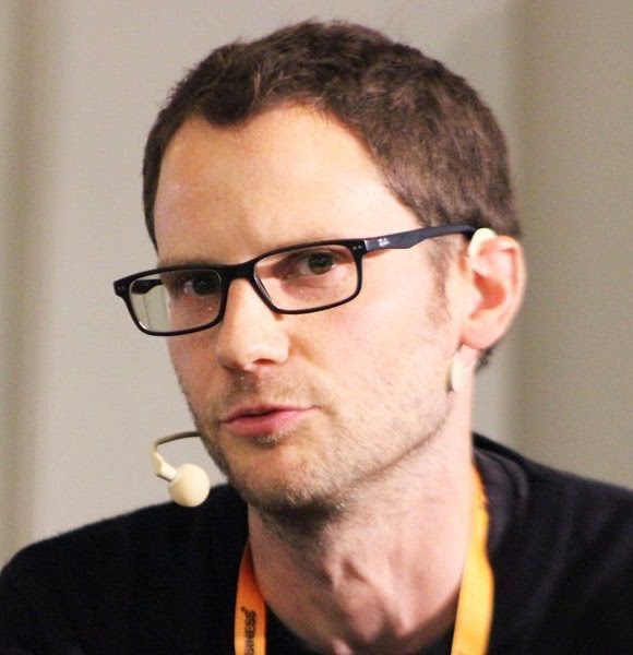
Dr. Ben Glocker
Reader in Machine Learning for Imaging at the Department of Computing at Imperial College London
Dr. Ben Glocker
Reader in Machine Learning for Imaging at the Department of Computing at Imperial College London
Biography: Ben Glocker is Reader in Machine Learning for Imaging at the Department of Computing at Imperial College London where he co-leads the Biomedical Image Analysis Group. He also leads the HeartFlow-Imperial Research Team and is scientific advisor for Kheiron Medical Technologies and a Visiting Researcher at Microsoft Research Cambridge. He holds a PhD from TU Munich and was a postdoc at Microsoft and a Research Fellow at the University of Cambridge. His research is at the intersection of medical imaging and artificial intelligence aiming to build computational tools for improving image-based detection and diagnosis of disease.
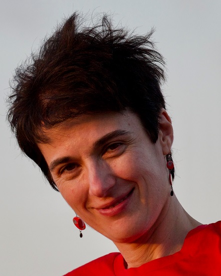
Prof. Aris Papageorghiou
Professor of Fetal Medicine and the Clinical Research Director of the Oxford Maternal and Perinatal Health Institute
Prof. Aris Papageorghiou
Professor of Fetal Medicine and the Clinical Research Director of the Oxford Maternal and Perinatal Health Institute
Biography: Aris is a Professor of Fetal Medicine and the Clinical Research Director of the Oxford Maternal and Perinatal Health Institute. He leads a number of research projects focused in the areas of maternal, fetal and perinatal health and using diverse methods - including basic science, clinical epidemiology, trials, knowledge transfer and implementation science. A major research interests is the use of artificial intelligence (AI) in pregnancy imaging and screening, and for several years he has worked with biomedical engineers on these problems. Through this work, Aris co-founded the Oxford University spin-out, Intelligent Ultrasound.
Important Dates
| Submission System Opens | January 2021 (Open now) |
| Paper Submission Deadline | |
| Author Notification (Regular Papers) | |
| Camera-ready regular papers due | |
| Conference Abstract Submission deadline | 31st May 2021 |
| Author Notification (Conference Abstracts) | 15th June 2021 |
| Camera-ready conference abstracts due | 22nd June 2021 |
| Conference | 12th - 14th July 2021 |
Call for Papers / Author Instructions
Consent for Publication
All accepted papers must fill and submit the consent to publish form. Please download and submit to CMT.
Camera-ready submission
All accepted papers must submit the following documents to CMT.
1- The paper in PDF format
2- The source of your paper. If the paper is written using MS Word, please submit the Word file. If the paper is written using LaTex, please compress all source files and submit one zip file.
3- The Consent for publication form (see above).
You are invited to submit your abstract paper to MIUA 2021 which will be held in Oxford, UK. The abstract paper submission system is now open.
The full paper submission deadline will be 23:59, Greenwich Mean Time (GMT), on 28th March 2021.
The abstract paper submission deadline will be 23:59, Greenwich Mean Time (GMT), on 31st May 2021.
High-quality papers are requested, containing original contributions to the topics within the scope of MIUA.
Paper Submissions:
For the 25th MIUA conference, we welcome submissions, as regular conference papers and conference abstracts.
Regular papers: Authors are invited to submit full papers of length between 8 and 15 pages (1 column – the LNCS template) showing original research contributions under the topics of the conference. All submissions will be double-blind peer-reviewed and accepted articles will be published as MIUA Proceedings by the Springer Publishing Group.
Conference abstracts: Authors are invited to submit short papers of length up to 3 pages excluding references (1 column – the LNCS template) showing proof-of-concept research contributions under the topics of the conference. All submissions will be peer-reviewed and accepted articles will be published as MIUA Abstract Proceedings on the MIUA website.
Accepted papers will be published in the MIUA proceedings in the Springer LNCS Series. Authors should consult Springer’s authors’ guidelines and use their proceedings templates, either for LaTeX or for Word, for the preparation of their papers. Springer encourages authors to include their ORCIDs in their papers. In addition, the corresponding author of each paper, acting on behalf of all of the authors of that paper, must complete and sign a Consent-to-Publish form. The corresponding author signing the copyright form should match the corresponding author marked on the paper. Once the files have been sent to Springer, changes relating to the authorship of the papers cannot be made.
MIUA continues to foster fairness, diversity, and inclusion within its community. Submissions from typically underrepresented groups are particularly encouraged.
Review Process
MIUA seeks your assistance as an expert reviewer for this annual conference. If you would like to review for MIUA, please contact us via: miua2021(at)eng.ox.ac.uk or twitter @miua2021. Please share this CfP with colleagues who might want to contribute to MIUA2021.
Registration Information
We are delighted to inform you that the registration fees will be reduced from normal registration fees for this year’s meeting.Student registration fee is £40
Non-student registration fee is £60
Please follow this link to register your attendance and your paper: Register now!.Conference Programme (all times are in GMT+1)
Access the conference proceeding
Monday 12th July 2021
| 09.00 – 09.15 | Welcome/opening remarks |
| 09.15 – 10.45 | Oral Session 1 (Biomarker Detection)
Session chairs: Michael Brady & Reyer Zwiggelaar 3 oral presentations: 10 min presentation each, followed by 15 minutes Q&A
3 oral presentations: 10 min presentation each, followed by 15 minutes Q&A
|
| 10.45 – 11.00 | Break |
| 11.00 – 12.00 | Keynote Lecture
By Prof Aris Papageorghiou Title: Ultrasound in Fetal and Maternal Health Session chair: Mohammad Yaqub |
| 12.00 – 13.30 | Oral Session 2 (Image Segmentation)
Session chairs: Heba Sailem & Carlos Reyes-Aldasoro 3 oral presentations: 10 min presentation each, followed by 15 minutes Q&A
3 oral presentations: 10 min presentation each, followed by 15 minutes Q&A
|
| 13.30 – 14.00 | Break |
| 14.00 – 15.00 | Oral Session 3 (Image enhancement, quality assessment, and data privacy)
Session chairs: Maria Valdes Hernandez & Claudia Lindner 4 oral presentations: 10 min presentation each, followed by 20 minutes Q&A
|
| 15.00 – 16.00 | Poster session and networking (individual rooms for each presenter of the day) |
| 16.00 – 17.00 | Workshop: by MathWorks, Title: 3-D Brain Tumour Segmentation using Deep Learning with MATLAB
Session chair: Bartek Papież |
| 17.00 – 17.05 | Day 1 closing |
Tuesday 13th July 2021
| 09.00 – 10.00 | Oral Session 4 (Image Registration and reconstruction)
Session chairs: Sharib Ali & Tryphon Lambrou 4 oral presentations: 10 min presentation each, followed by 20 minutes Q&A
|
| 10.00 – 10.15 | Break |
| 10.15 – 11.00 | Oral Session 5 (Biomarker detection)
Session chairs: Bogdan Matuszewski & Guang Yang 3 oral presentations: 10 min presentation each, followed by 15 minutes Q&A
|
| 11.00 – 11.15 | Break |
| 11.15 – 12.15 | Keynote Lecture
By Prof Mihaela van der Schaar Title: The Role of Imaging in Machine Learning for Healthcare Session chair: Alison Noble |
| 12.15 – 12.45 | Break |
| 12.45 – 14.00 | Oral Session 6 (Classification)
Session chairs: Harshita Sharma & Michael Brady 3 oral presentations: 10 min presentation each, followed by 15 minutes Q&A
2 oral presentations: 10 min presentation each, followed by 10 minutes Q&A
|
| 14.00 – 14.15 | Break |
| 14.15 – 14.45 | Lightning presentation for abstracts (pre-recorded)
|
| 14.45 – 15.45 | Poster session and networking (individual rooms for each presenter of the day) |
| 15.45 – 16.00 | Break |
| 16.00 – 17.00 | Workshop: by NVIDIA, Title: Optimising deep learning solutions with PyTorch and TensorFlow
Session chair: Ana Namburete |
| 17.00 – 17.05 | Day 2 closing |
Wednesday 14th July 2021
| 09.00 – 10.00 | Oral Session 7 (Image Registration and reconstruction)
Session chairs: Saad Jbabdi & Yalin Zheng 4 oral presentations: 10 min presentation each, followed by 20 minutes Q&A
|
| 10.00 – 10.15 | Break |
| 10.15 – 11.15 | Keynote Lecture
By Dr. Ben Glocker Title: Towards Safer AI in Medical Imaging Session chair: Ana Namburete |
| 11.15 – 12.15 | Poster session and networking (individual rooms for each presenter of the day) |
| 12.15 – 13.15 | Break |
| 13.15 – 14.30 | Oral Session 8 (Biomedical simulation and modelling (incl. generative models)
Session chairs: Robail Yasrab & Jens Rittscher 3 oral presentations: 10 min presentation each, 15 min QA for all papers
2 oral presentations: 10 min presentation each, 10 min QA for all papers
|
| 14.30 – 14.45 | Break |
| 14.45 – 15.30 | Oral Session 9 (Radiomics, predictive models, and quantitative imaging biomarkers)
Session chairs: Omar Al-Kadi & Xujiong Ye 3 oral presentations: 10 min presentation each, followed by 15 minutes Q&A
|
| 15.30 – 15.45 | Closing remarks, Prizes, and MIUA 2022 Presentation |
Our Sponsors
We gratefully acknowledge the generous support provided by the following patrons:
Premium Sponsorship
Basic Sponsorship
Basic Sponsorship

Basic Sponsorship
Awards
We gratefully acknowledge the generous support provided by the following patrons to sponsor the following awards:
Students best and runner up papers
Best paper award
Organizing Committee
The Organizing Committee is comprised of academic members from the Medical Sciences Division (Nuffield Department of Clinical Neurosciences [NDCN], and The Big Data Institute [BDI]) and the Mathematical Physical and Life Sciences Division (Institute of Biomedical Engineering [IBME]), representing Oxford’s core strategic partners in medical imaging research.
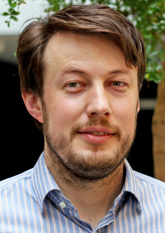
Bartlomiej Papiez
Research Fellow at the Big Data Institute.
Bartlomiej Papiez
BDI, Oxford
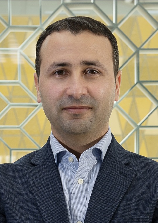
Mohammad Yaqub
Assistant Professor at MBZUAI (Abu Dhabi) and Research Fellow at NDCN (Oxford).
Mohammad Yaqub
MBZUAI, Abu Dhabi and NDCN, Oxford
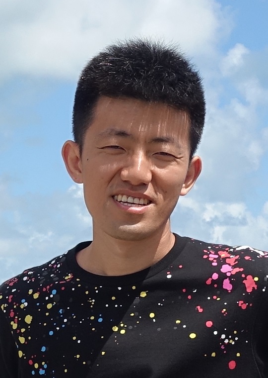
Jianbo Jiao
Researcher at the Biomedical Image Analysis (BioMedIA) group and the Visual Geometry Group (VGG), Engineering SCience.
Jianbo
Jiao
IBME & VGG, Oxford
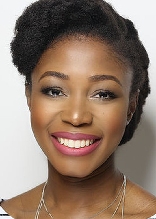
Ana Namburete
Royal Academy Research Fellow based at the Institute of Biomedical Engineering.
Ana
Namburete
IBME, Oxford
Alison Noble
Technikos Professor of Biomedical Engineering at the Institute of Biomedical Engineering.
Alison
Noble
IBME, Oxford
Contact Us
For further information, please contact us via email and we will get back to you as soon as we can.
Institute of Biomedical Engineering
Old Road Campus Research Building
Roosevelt Drive, Oxford
United Kingdom OX3 7DQ
miua2021@eng.ox.ac.uk
@miua2021